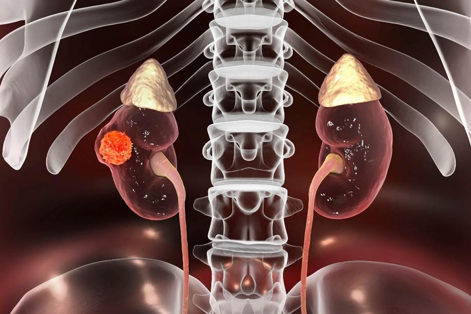

Hydronephrosis is often described simply as a “swollen kidney”, but the appearance of a dilated kidney on a scan is only the beginning of the clinical picture. The more important question is why the kidney is swollen, whether urine is able to drain effectively, and whether the underlying problem is beginning to threaten kidney function. […]
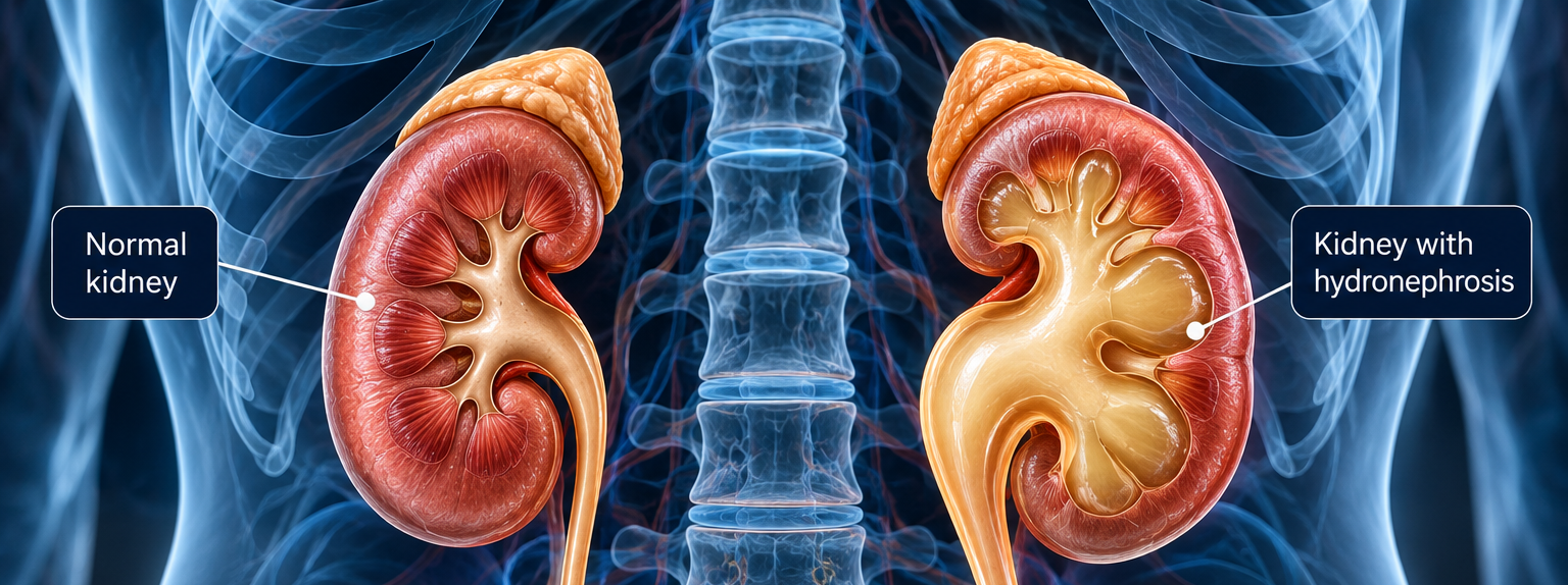
Hydronephrosis is often described simply as a “swollen kidney”, but the appearance of a dilated kidney on a scan is only the beginning of the clinical picture. The more important question is why the kidney is swollen, whether urine is able to drain effectively, and whether the underlying problem is beginning to threaten kidney function.
In some patients, hydronephrosis may be mild and require observation rather than immediate intervention. In others, it may indicate a significant urinary obstruction that needs treatment. When obstruction is severe, persistent, associated with infection, or begins to compromise kidney function, delaying treatment can have important consequences.
For the urologist, the objective is therefore not simply to make the swelling disappear on an ultrasound or CT scan. The priority is to understand what is happening to the kidney behind the obstruction and, where necessary, restore drainage before useful kidney function is permanently lost.
The kidneys continuously produce urine, which normally drains through the ureters into the bladder. When that drainage pathway is obstructed or significantly impaired, urine can accumulate above the affected area and cause the kidney’s collecting system to dilate. This dilation is known as hydronephrosis.
Hydronephrosis tells us that the urinary drainage system is dilated, but it does not tell us the underlying cause or, by itself, how much kidney function has been affected.
There are many possible causes. A stone may become lodged within the ureter, scar tissue may cause a narrowing or stricture, or there may be an obstruction where the kidney joins the ureter, known as ureteropelvic junction (UPJ) obstruction. Previous surgery or injury can alter the urinary tract, while in some patients an abdominal or pelvic process may compress the ureter from outside.
This is why the discovery of hydronephrosis needs to be interpreted in context. The important clinical question is not simply whether hydronephrosis is present, but what is causing it and what effect it is having on the kidney.
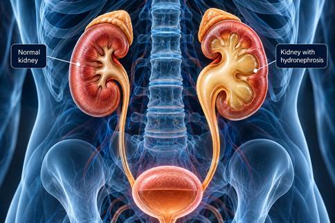
When urine cannot pass normally through the urinary tract, pressure can build upstream from the obstruction. The collecting system of the kidney begins to dilate as urine accumulates, producing the appearance of hydronephrosis on imaging.
If the obstruction is temporary or relieved sufficiently early, kidney function may be preserved or may recover. However, persistent obstruction can place continued pressure on the kidney and eventually interfere with its ability to function normally.
The relationship between obstruction and kidney damage is not identical in every patient. The severity and duration of the obstruction, whether one or both kidneys are affected, the patient’s underlying kidney health and the presence of infection can all influence the clinical significance.
This is also why the degree of swelling visible on a scan should not be interpreted in isolation. A markedly dilated kidney does not automatically mean that the kidney has lost all useful function, while a less dramatic radiological appearance does not necessarily mean that an obstruction can safely be ignored.
The anatomy and the function need to be considered together.
One of the misconceptions surrounding urinary obstruction is that a serious problem will always cause severe pain.
An acutely obstructing kidney stone can certainly produce intense flank pain, which is why many people associate urinary obstruction with a dramatic presentation. However, obstruction can also develop gradually. In these circumstances, symptoms may be intermittent, relatively mild or sometimes absent.
Some patients may experience an ache in the flank, recurrent urinary infections, nausea or episodes of discomfort that come and go. In other cases, hydronephrosis may be discovered unexpectedly during imaging performed for an unrelated reason.
The absence of severe pain therefore does not necessarily establish that the kidney is safe.
When hydronephrosis is identified, the assessment needs to determine whether there is a genuine obstruction and, importantly, whether that obstruction is affecting drainage or renal function.
There is rarely a single investigation that answers every question about an obstructed kidney. Instead, the clinical history, symptoms, imaging and kidney function are considered together.
Ultrasound can demonstrate dilation of the kidney’s collecting system and is frequently useful for identifying and monitoring hydronephrosis. CT imaging can provide considerably more anatomical information and may help identify a stone, narrowing, surrounding abnormality or another possible cause of obstruction.
Blood tests can provide information about overall kidney function, but they also have limitations. When the opposite kidney is functioning normally, overall blood results may remain relatively reassuring even when one kidney is experiencing a significant problem.
In selected patients, a nuclear medicine renal scan or renogram can provide additional information about how each kidney is functioning and how effectively urine drains from it.

This distinction is important. Imaging can tell the surgeon a great deal about what the urinary tract looks like, while functional assessment can help answer a different question: how well is the kidney actually working?
The decision to intervene is therefore not based on hydronephrosis alone. The cause of the obstruction, symptoms, infection risk, drainage, remaining kidney function and evidence of deterioration all contribute to the decision.
Not every patient with hydronephrosis requires emergency treatment. There are, however, circumstances in which urinary obstruction becomes significantly more concerning.
One of the most important is the combination of obstruction and infection. When an infected urinary system is obstructed, infected urine may be unable to drain normally. This can become a serious clinical situation requiring urgent medical assessment and, in appropriate cases, decompression of the urinary system.
Deteriorating kidney function is another important concern because it may indicate that obstruction is no longer simply an anatomical finding but is beginning to have a functional consequence.
The situation also requires particular consideration when both kidneys are obstructed or when obstruction affects a solitary functioning kidney. In these circumstances, there may be considerably less renal reserve available if drainage becomes compromised.
Severe or persistent symptoms, recurrent infections and evidence that hydronephrosis or renal function is worsening can similarly influence how quickly intervention is required.
The urgency therefore comes from the effect of the obstruction, rather than from the word “hydronephrosis” itself.
When a kidney is threatened by obstruction, the immediate objective is not always to perform the final reconstructive operation.
Sometimes the first priority is to relieve the pressure and allow urine to drain.
Depending on the clinical circumstances, temporary drainage may be achieved with a ureteric stent, which provides an internal pathway for urine to pass from the kidney to the bladder, or with a nephrostomy tube, which drains the kidney externally through the back.
These interventions do not necessarily correct the underlying cause. Instead, they can provide a way to decompress the urinary system while infection is treated, kidney function is reassessed, or definitive treatment is planned.
This distinction between draining the kidney and correcting the obstruction is important.
In an urgent situation, protecting the kidney and the patient comes first. The definitive reconstruction can then be considered once the immediate problem has been stabilised.
Once the cause of hydronephrosis has been established, treatment needs to address that specific problem.
A ureteric stone requires a different approach from a ureteric stricture. An obstruction caused by scar tissue after previous surgery may require a different strategy from a congenital UPJ obstruction. External compression of the ureter introduces another set of considerations entirely.
For UPJ obstruction, laparoscopic or robotic pyeloplasty may be used to remove or bypass the narrowed segment and reconstruct the drainage pathway between the kidney and ureter. In more complex cases involving the ureter, previous operations, scarring or loss of healthy tissue, reconstructive surgery may require a more individualised approach.
This is where the objective extends beyond simply “removing a blockage”.
The surgeon needs to establish a drainage pathway that works while preserving the healthy anatomy and kidney function that remain.

Urinary obstruction can become particularly complex when the patient has already undergone surgery in the same area.
Scar tissue may distort normal anatomical planes, a ureter may have been previously repaired or repositioned, and the amount of healthy tissue available for another reconstruction may be different from what would be expected during a first operation.
In these circumstances, the surgeon needs to understand not only where the obstruction is located, but also what has happened to the surrounding anatomy before. Previous operative reports, imaging and information about earlier complications can therefore become important components of surgical planning.
Revision surgery is not simply an attempt to repeat the first procedure. The reconstruction needs to be designed around the anatomy and function that exist now.
One of the more difficult situations occurs when obstruction has been present long enough for the affected kidney to lose some of its function.
At this point, finding the obstruction is only part of the assessment. The surgeon also needs to determine how much useful function remains, whether that function may benefit from restored drainage and what treatment is appropriate for the individual patient. This is where functional assessment becomes particularly valuable.
The objective of reconstructive surgery is not simply to produce a urinary tract that appears less dilated on imaging. A kidney may remain somewhat dilated even after drainage has improved, particularly when hydronephrosis has been longstanding.
The more meaningful objective is to preserve useful renal function and establish effective drainage where this is achievable. This distinction is important because successful treatment should not be judged purely by how the kidney looks on a scan.
Hydronephrosis should be understood as a sign that requires an explanation rather than as a diagnosis in itself.
For some patients, careful observation and follow-up may be appropriate. For others, the underlying cause needs treatment. When obstruction is accompanied by infection, worsening kidney function or other high-risk circumstances, restoring drainage can become considerably more urgent.
The central surgical question is therefore not simply, “How swollen is the kidney?”
It is whether there is a meaningful obstruction, what is causing it, how well the kidney is functioning, whether that function is changing and what needs to be done to protect it.
When intervention becomes necessary, treatment may range from temporary drainage to definitive minimally invasive or reconstructive surgery, depending on the underlying problem.
Ultimately, the purpose of treating urinary obstruction is not simply to correct an abnormality visible on a scan. It is to protect the functioning kidney behind it before potentially recoverable function becomes permanently lost.