
Being told that one of your kidneys has reduced function can raise some difficult questions. Does this mean the kidney is failing? Can its function improve? Will an operation be necessary? And perhaps most importantly, is the kidney still worth preserving? These questions cannot usually be answered by one scan result alone. Kidney function needs […]
Being told that one of your kidneys has reduced function can raise some difficult questions. Does this mean the kidney is failing? Can its function improve? Will an operation be necessary? And perhaps most importantly, is the kidney still worth preserving?
These questions cannot usually be answered by one scan result alone. Kidney function needs to be understood in the context of the patient’s symptoms, anatomy, medical history and the underlying reason for the reduction in function.
In urology, the challenge is not simply identifying that a kidney is functioning less effectively. It is understanding why that has happened, whether the problem is continuing and what can reasonably be done to protect the remaining function.
The phrase “reduced kidney function” can refer to different findings, and understanding that distinction is important.
Blood tests, including creatinine measurements and estimated glomerular filtration rate (eGFR), provide information about overall kidney function. However, they do not necessarily tell us how much work each individual kidney is performing.
When both kidneys are present, one kidney may compensate for reduced function in the other. As a result, a patient’s overall blood results can sometimes appear relatively reassuring even when one kidney has a significant problem.
This is one reason why a urologist may recommend additional investigations to understand the contribution and drainage of each kidney individually.
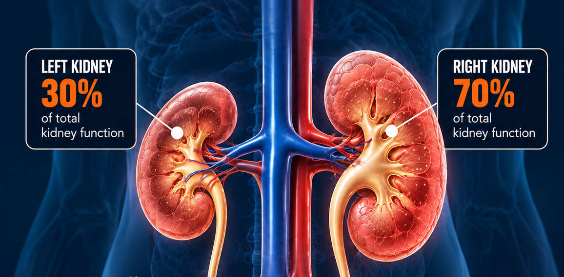
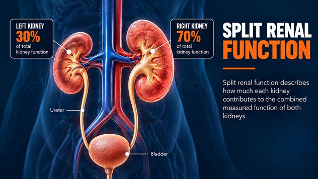
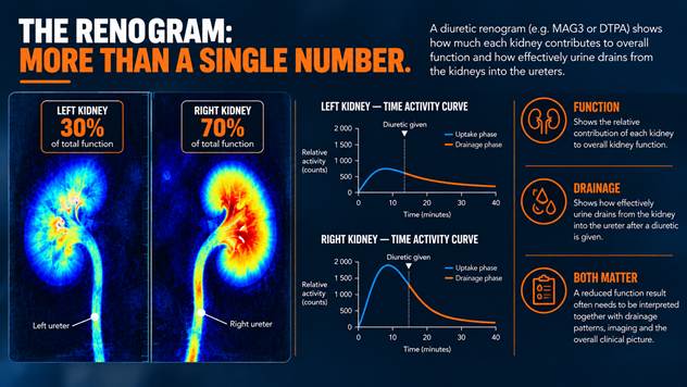
Split renal function, also called differential renal function, describes the relative contribution of each kidney to the combined function measured during a nuclear medicine renal study.
In a relatively balanced result, each kidney may contribute approximately half of the measured total. However, this distribution is not always equal.
For example, a scan may indicate that one kidney contributes 30% and the other contributes 70%.
This does not mean that the first kidney has exactly 30% of its original function remaining. It means that, during the investigation, it accounted for approximately 30% of the measured combined renal contribution.
That distinction matters because split renal function is a relative measurement, not a complete description of the kidney’s absolute functional capacity.
A lower percentage is therefore an important finding, but it does not independently establish whether the kidney can recover, whether surgery is required or whether the kidney should be removed.
A renal nuclear medicine scan, often referred to as a renogram, can help clinicians assess how the kidneys take up and process a tracer and how effectively it passes through the urinary collecting systems.
In selected cases, a diuretic may be administered during the investigation to help assess urinary drainage.
The results can provide valuable information about the relative contribution of each kidney and whether drainage appears delayed.
However, interpreting a renogram requires more than looking at a percentage or a drainage curve. The patient’s hydration, bladder emptying, kidney function, anatomy and the technical circumstances of the study can influence the findings.
A kidney with substantially reduced function can also be more difficult to assess accurately for obstruction.
For these reasons, the renogram is considered alongside other investigations rather than treated as a standalone answer.
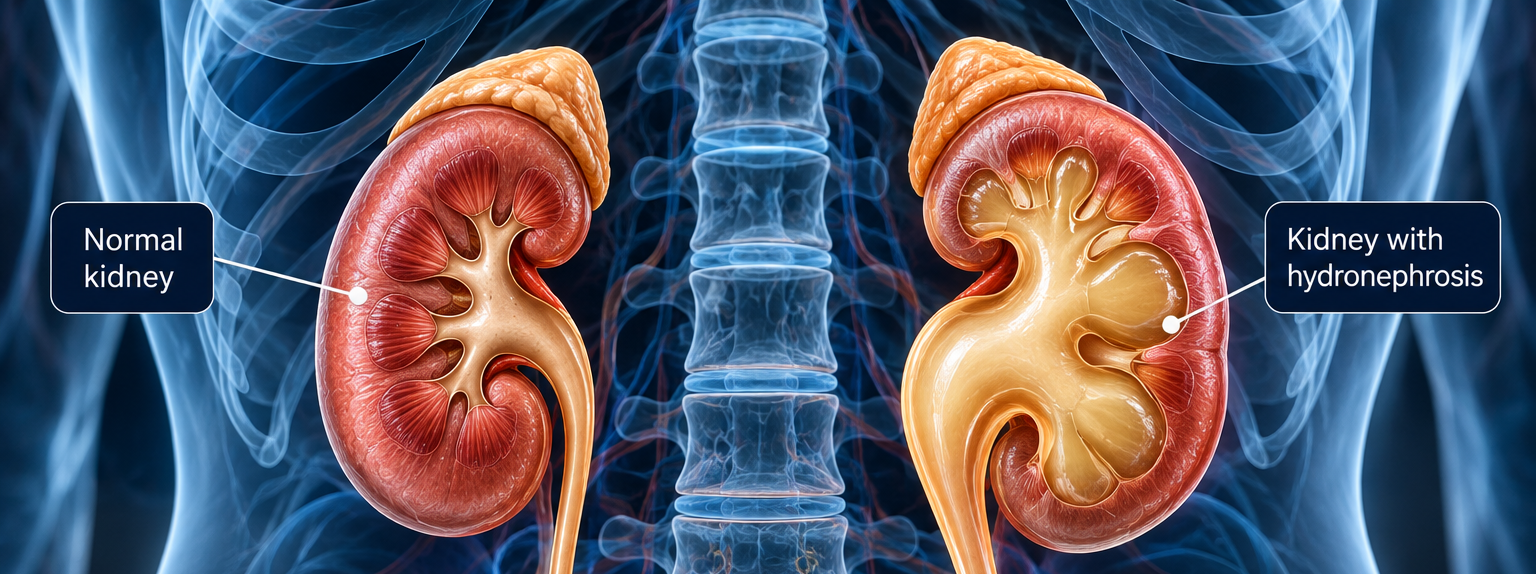
Normal kidney function depends partly on urine being able to drain through the urinary tract without significant obstruction.
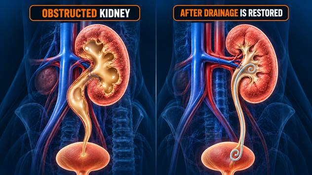
When an obstruction develops, pressure may increase within the collecting system. If the obstruction is sufficiently severe or persists for long enough, it can interfere with normal kidney function and potentially cause lasting damage.
There are several possible causes, including ureteric stones, narrowing of the ureter, ureteropelvic junction obstruction and anatomical changes associated with previous surgery.
The consequences depend on the severity and duration of the obstruction, whether infection is present and the condition of the kidney before the problem developed.
Some impairment may improve when drainage is restored, particularly when the obstruction is addressed before irreversible damage develops. In other cases, longstanding injury may limit how much function can recover.
The important point is that relieving an obstruction and restoring lost kidney function are related goals, but they are not the same outcome.
When a kidney has reduced function, the decision is not simply between operating and doing nothing.
The surgeon must first understand what is causing the problem and whether that cause can be corrected.
Imaging can help establish the anatomy of the urinary tract, identify narrowing or obstruction and assess structural changes. Functional investigations can provide additional information about the kidney’s contribution and drainage.
The patient’s symptoms, recurrent infections, pain, overall renal function and the condition of the opposite kidney must also be considered.
Another important question is whether there is evidence that the affected kidney’s function is stable or continuing to deteriorate.
In selected patients, restoring urinary drainage may provide an opportunity to preserve useful remaining function. This could involve temporary drainage, treatment of a stone or reconstructive surgery to correct a narrowing or obstruction.
In more complex cases, surgical planning may involve determining whether a minimally invasive reconstructive procedure can provide a durable drainage pathway while preserving healthy tissue.
The objective is not simply to perform an operation because a scan is abnormal. It is to determine whether an intervention offers a meaningful benefit to the patient.
A particularly difficult question arises when a scan indicates that one kidney contributes only a small proportion of the combined function.
Although very low split renal function can influence treatment decisions, there is no single percentage that automatically determines the correct course of action for every patient.
Some kidneys with low function may still provide useful contribution, and certain patients may benefit from attempts to relieve obstruction or preserve the kidney.
In other circumstances, a severely damaged kidney may have little prospect of meaningful recovery, particularly when there are additional problems such as recurrent infections, persistent pain or other complications.
The decision must therefore consider the whole clinical picture, including the expected benefits and risks of preservation, reconstruction or removal.
This is especially important when the opposite kidney is also affected or when the patient’s overall renal reserve is limited.
Kidney function does not always deteriorate in a way that produces obvious symptoms. Some patients experience significant pain, while others have relatively few symptoms despite important abnormalities.
When reduced function is identified, timely investigation can help establish whether the condition is stable, whether there is an ongoing threat and whether treatment may prevent further deterioration.
For patients with complex urinary obstruction, previous surgery or altered anatomy, this assessment can require particularly careful planning.
In Dr MC Conradie’s field of minimally invasive and reconstructive urology, understanding the relationship between anatomy, obstruction and remaining kidney function is central to evaluating appropriate surgical options.
The aim is to make a considered decision based on the kidney’s current condition, the likely cause of the problem and the realistic possibilities for preserving useful function.
A scan showing reduced kidney function provides an important piece of clinical information, but it does not tell the entire story.
The next step is understanding what that finding means for the individual patient.
Is there an obstruction? Is the kidney still contributing useful function? Is that function stable? Can the underlying problem be corrected? And would intervention offer a meaningful opportunity to preserve the kidney?
These are the questions that guide the investigation and treatment process.
The goal is not simply to respond to a percentage on a scan. It is to make the most appropriate decision for the kidney and the patient.
For patients with reduced kidney function associated with suspected urinary obstruction or complex anatomical problems, specialist urological assessment can help determine the appropriate next steps.