
The kidneys play an essential, silent role in keeping the body in balance — filtering waste, maintaining electrolyte levels, and regulating blood pressure. When urine cannot drain properly from the kidney due to an obstruction at the ureteropelvic junction (UPJ), it can lead to pain, infections, and progressive kidney damage. For patients facing this condition, […]

The kidneys play an essential, silent role in keeping the body in balance — filtering waste, maintaining electrolyte levels, and regulating blood pressure. When urine cannot drain properly from the kidney due to an obstruction at the ureteropelvic junction (UPJ), it can lead to pain, infections, and progressive kidney damage.
For patients facing this condition, laparoscopic pyeloplasty offers a highly effective, minimally invasive solution that preserves the kidney while restoring normal function.
Understanding the Ureteropelvic Junction (UPJ) Obstruction
The ureteropelvic junction is the connection point where the kidney’s drainage system (the renal pelvis) joins the ureter — the narrow tube that carries urine to the bladder. In some patients, this junction becomes narrowed, kinked, or blocked, slowing or completely stopping the flow of urine.
When this occurs, urine begins to accumulate inside the kidney, leading to hydronephrosis (swelling of the kidney). Over time, this pressure can compress delicate kidney tissue, impairing its ability to filter blood and eventually leading to loss of kidney function.
Causes of UPJ Obstruction
UPJ obstruction may be:
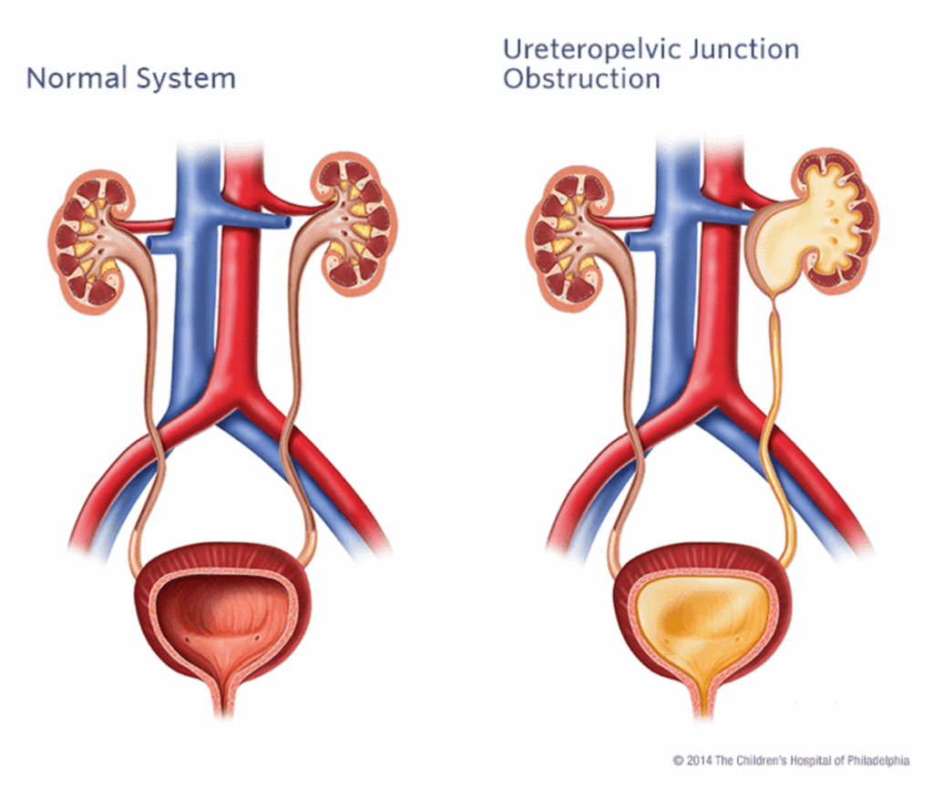
(Image Source: Children’s Hospital of Philadelphia, Link: https://www.chop.edu/conditions-diseases/ureteropelvic-junction-upj-obstruction )
Symptoms and Early Warning Signs
Because UPJ obstruction affects how urine drains, symptoms may vary depending on severity and cause. Common symptoms include:
In some patients, particularly children or those with mild obstruction, the condition may be discovered incidentally during imaging for another reason.
Diagnosing UPJ Obstruction: Functional and Anatomical Assessment
Accurate diagnosis is critical for choosing the correct treatment approach. At Dr MC Conradie Inc., diagnosis involves a combination of anatomical imaging and functional studies to determine the site and severity of the blockage:
Each patient’s results are carefully reviewed to confirm the diagnosis and plan a tailored surgical strategy.
When Is Surgery Needed?
Mild obstruction may sometimes be monitored if kidney function remains stable. However, surgery becomes necessary when:
At this point, laparoscopic pyeloplasty is considered the gold standard for durable, anatomical correction.
What Is Laparoscopic Pyeloplasty?
Laparoscopic pyeloplasty is a minimally invasive reconstructive procedure that removes the narrowed or obstructed section of the ureter and reattaches the healthy ends — restoring a smooth, open connection between the kidney and ureter.
Instead of the large incision required for open surgery, this approach uses three to four small incisions through which a camera and fine instruments are inserted. The entire procedure is performed under magnified vision, allowing precise dissection and delicate suturing.
A temporary stent (a thin internal tube) is placed between the kidney and bladder to support healing and ensure continuous drainage during recovery.
Advantages of the Laparoscopic Approach
Compared with open surgery, laparoscopic pyeloplasty offers substantial benefits:
Studies consistently show success rates of over 90–95% when performed by experienced laparoscopic surgeons.
Comparing Treatment Options: Laparoscopic vs. Endopyelotomy vs. Open Surgery
When managing UPJ obstruction, several treatment methods exist — but their outcomes and indications differ significantly.
| Approach | Description | Advantages | Limitations | Best For |
| Endopyelotomy | An internal incision made endoscopically to widen the narrowed area. | Minimally invasive, short hospital stay. | Lower success (60–80%), high recurrence, not suitable for long strictures or crossing vessels. | Mild obstructions, high-risk surgical patients. |
| Laparoscopic Pyeloplasty | Precise excision of narrowed segment with anatomical reconstruction. | Gold standard success rates (>90%), long-term correction, minimal invasiveness. | Technically demanding; requires advanced laparoscopic expertise. | Most patients with primary or secondary UPJ obstruction. |
| Open Pyeloplasty | Traditional surgery via large flank incision. | Durable results, allows tactile feedback. | More pain, scarring, and recovery time; longer hospitalisation. | Complex redo cases or limited access to laparoscopic surgery. |
In skilled hands, laparoscopic pyeloplasty offers the best balance of long-term success, cosmetic outcome, and patient comfort — now considered the benchmark standard for UPJ repair worldwide.
Dr MC Conradie’s Expertise in Upper Tract Reconstruction
Dr MC Conradie is one of South Africa’s foremost urological surgeons in laparoscopic reconstructive surgery. Her expertise extends across complex renal and ureteral procedures, with a strong focus on organ preservation and functional restoration.
She has extensive experience in performing laparoscopic pyeloplasty for both congenital and acquired UPJ obstruction — combining meticulous surgical precision with an understanding of each patient’s anatomy and long-term health goals.
Every procedure is carefully planned using advanced imaging, and surgical techniques are adapted to the patient’s unique anatomy — whether due to a crossing vessel, fibrosis, or congenital narrowing.
Recovery and Follow-Up
After laparoscopic pyeloplasty:
The results are typically long-lasting, with improved pain control, restored drainage, and stabilisation or improvement in kidney function.
When to See a Specialist
Patients experiencing chronic flank pain, recurrent infections, or evidence of kidney swelling should seek prompt evaluation from a urologist specialising in minimally invasive surgery.
Early diagnosis and intervention can prevent irreversible kidney damage and avoid the need for complete nephrectomy (kidney removal).
At Dr MC Conradie Inc., patients receive comprehensive evaluation and world-class surgical care — with the goal of restoring normal flow and preserving every possible nephron.
Restoring Flow. Preserving Function. Improving Quality of Life.
Laparoscopic pyeloplasty is not simply about removing an obstruction — it’s about restoring the natural balance of the urinary system. With unmatched expertise and dedication to minimally invasive excellence, Dr MC Conradie provides patients with the highest standard of care, helping them regain comfort, confidence, and kidney health.

